Anatomy Of Upper Thigh And Hip / Gluteal and Psoas Relationship for Yogis | Love Yoga Anatomy : The hip's unique anatomy enables it to be both extremely strong and amazingly flexible, so it can bear weight and allow for a wide range of movement.
Anatomy Of Upper Thigh And Hip / Gluteal and Psoas Relationship for Yogis | Love Yoga Anatomy : The hip's unique anatomy enables it to be both extremely strong and amazingly flexible, so it can bear weight and allow for a wide range of movement.. Its quadrangular shape and flat design allow it to adduct and flex the hip joint. It is part of the lower limb. In vertebrate anatomy, hip (or coxa in medical terminology) refers to either an anatomical region or a joint. Chief flexor of knee weak. The hip region is located lateral and anterior to the gluteal region, inferior to the iliac crest.
Quadriceps, a group of four. Want to learn more about it? Superficial fascia.—the superficial fascia forms a continuous layer over the whole of the thigh; B, muscles of the anterior thigh compartment. In clinical anatomy the thigh muscles are divided into three groups:
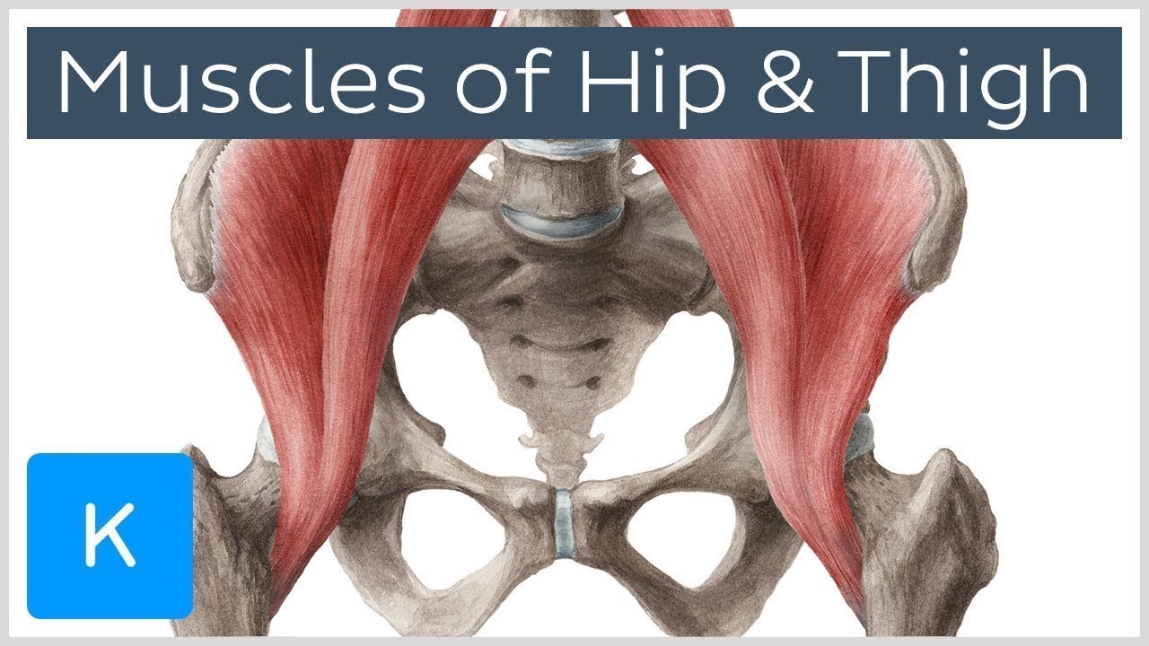
Muscles of the Hip and Thigh - Human Anatomy | Kenhub ... from i.ytimg.com Mri of upper leg (femur). The femur or thigh bone is one of the longest bones in the human body. Groin, inguinal region and the anterior and posterior regions of the hip and thigh. Each pelvic girdle consists of a hip bone (coxal bone, innominate bone), which articulates with the head of a femur. Jew anatomy atlases, the anatomy atlases logo, and a digital library of anatomy information are all trademarks of michael p. The femur, the hip bone (subdivided into ilium. Superficial fascia.—the superficial fascia forms a continuous layer over the whole of the thigh; Hip and knee pain and hip and shoulder pain are.
The different anatomical areas of the gluteal region:
The patient lies supine with the hip and knee flexed and the hip externally rotated into the frog leg position. May 13, 2019 edited by dr. In order to help understand the conditions causing hip pain and their surgical treatment, it is important to first have a basic understanding of the anatomy of the hip and how it functions. Hip anatomy, hip joint, groin anatomy. Anatomy hip, thigh and leg muscles. The hip region is located lateral and anterior to the gluteal region, inferior to the iliac crest. Quadriceps, a group of four. It is referred to as a ball and socket joint, and is. Each pelvic girdle consists of a hip bone (coxal bone, innominate bone), which articulates with the head of a femur. Want to learn more about it? Medial condyle of tibia nerve supply: The iliopsoas muscle, which extends from the lower back to upper femur; The body part defined by the hip joint and surrounding structures, including the region from the iliac crest to the thigh.
The patient lies supine with the hip and knee flexed and the hip externally rotated into the frog leg position. The femur, the hip bone (subdivided into ilium. for detailed anatomy of pelvic bones, read anatomy of hip bone. In vertebrate anatomy, hip (or coxa in medical terminology) refers to either an anatomical region or a joint. The thigh is the area between the hip and the knee joint.
Illustrations in Collection of Medical Illustrations ... from www.netterimages.com Unlike the shoulder girdle, the pelvic girdle is firmly integrated into the axial skeleton: Knowing the anatomy of your hip can help you understand the source of any hip pain. The uppermost of the medial thigh muscles is the pectineus muscle. The iliopsoas muscle, which extends from the lower back to upper femur; Tibial part of the sciatic nerve action: The patient lies supine with the hip and knee flexed and the hip externally rotated into the frog leg position. Quadriceps, a group of four. Iliopsoas muscle, a hip flexor muscle that attaches to the upper thigh bone.
The body part defined by the hip joint and surrounding structures, including the region from the iliac crest to the thigh.
The iliopsoas muscle, which extends from the lower back to upper femur; It functions to adduct the thigh and to flex and rotate the leg medially at the knee. Hip movements include flexion, extension, abduction, adduction, circumduction, and hip rotation. The upper part of the thigh bone consists of the femoral head, femoral. Unlike the shoulder girdle, the pelvic girdle is firmly integrated into the axial skeleton: B, muscles of the anterior thigh compartment. The paired hip bones are connected. Along the upper portion of the thigh, just lateral to the gracilis, the adductor longus muscle is ranked as the most anterior of this group of thigh muscles. Groin, inguinal region and the anterior and posterior regions of the hip and thigh. While the thigh muscles will be slip into the anterior, medial and posterior groups. Knee assessment and hip mechanics learn how hip and pelvis mechanics can influence the knee powered by physiopedia start course. Related online courses on physioplus. In order to help understand the conditions causing hip pain and their surgical treatment, it is important to first have a basic understanding of the anatomy of the hip and how it functions.
Knee assessment and hip mechanics learn how hip and pelvis mechanics can influence the knee powered by physiopedia start course. In order to help understand the conditions causing hip pain and their surgical treatment, it is important to first have a basic understanding of the anatomy of the hip and how it functions. Chief flexor of knee weak. The single bone in the thigh region is called the origin: Asymmetrical gluteal or thigh skin folds.
Left hip pain - not sure if serious from imbuebody.com Groin, inguinal region and the anterior and posterior regions of the hip and thigh. This deep muscle begins in the low back and pelvis and connects on the inside edge of the upper femur. Here are a few fundamental moves to try. The femur, the hip bone (subdivided into ilium. Hip surgeon dr guillaume dumont offers hip pain treatments in columbia, sc. Hip flexor deep in pelvis a composite o… used to extend the hip when climbing st… Hip movements include flexion, extension, abduction, adduction, circumduction, and hip rotation. The patient lies supine with the hip and knee flexed and the hip externally rotated into the frog leg position.
Twists the leg out and twists the knee in toward your other leg.
Want to learn more about it? Here are a few fundamental moves to try. The uppermost of the medial thigh muscles is the pectineus muscle. The femur or thigh bone is one of the longest bones in the human body. Anatomy of the human body. Bones of the lower limb. Unlike the shoulder girdle, the pelvic girdle is firmly integrated into the axial skeleton: The paired hip bones are connected. Twists the leg out and twists the knee in toward your other leg. Superficial fascia.—the superficial fascia forms a continuous layer over the whole of the thigh; 340 anatomical structures of the hip region were labeled, accessible on anatomical parts: The thigh is the area between the hip and the knee joint. Asymmetrical gluteal or thigh skin folds.
Anatomy hip, thigh and leg muscles upper thigh anatomy. Sartorius muscle anatomy page has origin, insertion, innervation, and blood supply information.


0 Komentar